Brain function/structure measurements is rapidly improving due to the advancements in neuroimaging technologies. However, despite these improvements it is becoming increasingly necessary to make sense of the obtained high dimensional datasets which qualify to be called “big data”. Both univariate and multivariate methods have been popular in neuroimaging.
A univariate neuroimaging approach can identify individual voxels which differ between two groups (depicted as red dots in Figure 1), but their inter-relationships are not identified. Multivariate approaches allow relationships between the varying voxels to be captured (depicted as clusters of different colored dots IN Figure 1). Multivariate methods like source based morphometry (SBM) combines information across different voxels to identify covarying ‘networks,’ and performs testing on subject covariation of these networks rather than testing each voxel separately between two groups (Xu, Groth, et al., OHBM 2009; C.N.Gupta et al, Schizophrenia Bulletin, 2014). SBM identifies patterns of common variation (e.g. gray or white matter patterns) among subjects and preserves spatial correlation between different brain regions. Multivariate methods have several advantages over voxel or region of interest-based approaches as it 1) does not require a priori selection of regions to analyze and 2) acts as a spatial filter to separate signals which overlap but have different behavior (e.g. artifacts vs real changes in brain structure).

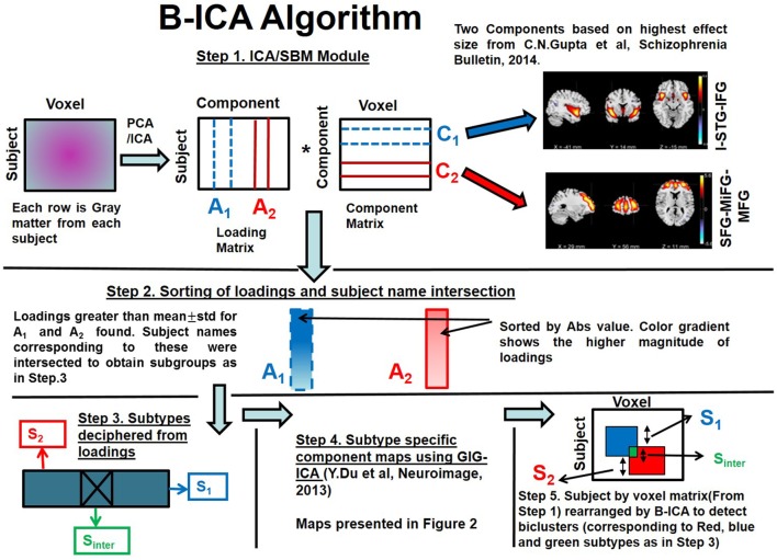
Biclustered independent component analysis is a multivariate clinical subtyping method applied to schizophrenia only cohort using structural MRI data. This framework includes both SBM and biclustering modules (Gupta et al., Frontiers in Psychiatry, 2017). This work exploited the joint distribution of the loadings to obtain subtypes, which were then tested for association with clinical symptoms as illustrated. Varying directionalities of GMC were observed in this work among the three homogeneous subtypes. The subtypes were obtained by considering loadings from two components comprising regions of insula/superior temporal gyrus/inferior frontal gyrus (I-STG-IFG component) and superior/medial/middle frontal gyrus (SFG-MiFG-MFG component) which had high effect size and showed diagnostic differences (Gupta et al., Frontiers in Psychiatry, 2017).
The goal of multimodal fusion is to capitalize on the strength of each modality in a joint analysis, rather than performing a separate analysis (J Sui et al, J Neurosci Methods,2012).Parallel ICA is a framework to investigate the integration of data from two imaging modalities, this method identifies independent components of both modalities and connections between them through enhancing intrinsic interrelationships (J.Liu et al, IEEE Signal Process Letters,2008; C.N.Gupta et al, Frontiers in Human Neuroscience, 2015). Recently, parallel independent component analysis (P-ICA) was applied to find connections between white matter of brain with genetic data for healthy controls and patients with schizophrenia.The FA component reflected decreased white matter integrity in the forceps major for SZ patients. The SNP component was overrepresented in genes whose products are involved in corpus callosum morphology (e.g., CNTNAP2, NPAS3, and NFIB) as well as canonical pathways of synaptic long term depression and protein kinase A signaling.
[1] Xu, Groth, et al., OHBM 2009; C.N.Gupta et al, Schizophrenia Bulletin, 2014
[2] Gupta, Cota Navin, et al. "Biclustered independent component analysis for complex biomarker and subtype identification from structural magnetic resonance images in schizophrenia." Frontiers in psychiatry 8 (2017): 179.